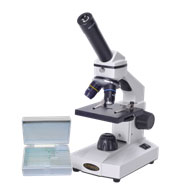
Source: Compound Microscope, Microscope.com
In order to study cells, scientists use microscopes. You have probably used microscopes similar to the one pictured here in science class.

Source: Compound Microscope, Microscope.com
Scientists use large electron microscopes, like the ones pictured below, in microscopy. Electron microscopes can magnify objects up to 10,000,000 times!
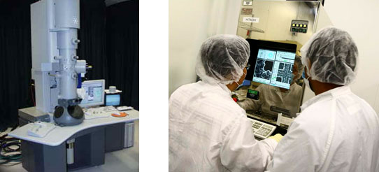
Source: Transmission Electron Microscope, The Chinese University of Honk Kong
Hitachi S-7800 SEM, Arizona State University
The graphic below shows what you can see with different types of microscopes. With the use of microscopes, it is possible to study everything from cells, viruses, single molecules or even atoms!
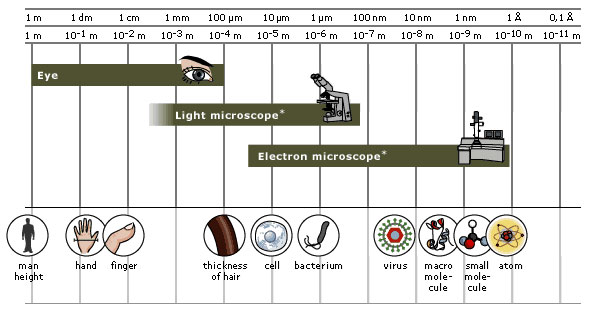
Source: Resolving Power of Microscopes, Nobel Prize.Org
Microscopes have come a long way since the first one built in late 16th century.

Source: Early microscopes, Molecular Expressions
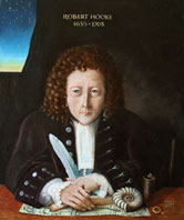
Source: Portrait of Robert Hooke, Wikimedia Commons
Robert Hooke, a scientist, discovered the cell. In 1665, he observed thin slices of cork from a cork tree under a microscope.
Hooke observed empty spaces contained by walls that he described as tiny boxes or a honeycomb. He called the structures cells because they reminded him of the rooms in a monastery. What Hooke saw was actually the cell walls of the plant cells. At that point he had no idea about the function of cells or that they contained other organelles.
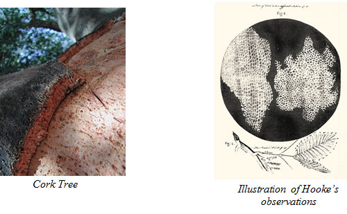
Source: Cork Tree, The Rare Pair
Robert Hooke's Observation of Cells, Wikimedia Commons
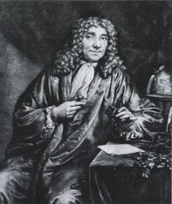
Source: Anton van Leeuwenhoek, Wikimedia Commons
In 1674, Antony van Leeuwenhoek was the first person to view and describe live cells under a microscope. Leeuwenhoek named the moving organisms that he saw animalcules, meaning "little animals.”
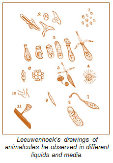
Source: Leeuwenhoek’s Animalcules, Internetlooks.com
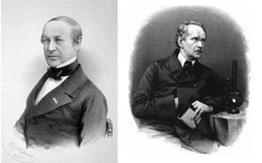
Source: Theodor Schwann and Matthias Jacob Schleiden, Wikimedia Commons
Two German scientists named Mattias Schleiden and Theodor Schwann continued the work of Hooke and Leeuwenhoek. In 1838, their work was published and their observations were summarized into the following three conclusions:
Today, Schleiden and Schwann’s first two observations are known to be correct but their third statement is incorrect.
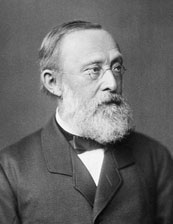
Source: Rudolf Virchow, Wikimedia Commons
Rudolf Virchow was a German physician who studied cell pathology, or the study of the nature of disease and its causes, concluded that cells must arise from pre-existing cells. In 1858, he published his findings and the cell theory was changed to the following:
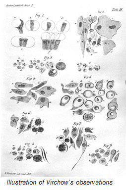
Source: Virchow-cell, Wikimedia Commons
As technology continued to develop, and scientists learned more about the structure and functions of cells, the cell theory was added to. The modern cell theory states the following: